
Spinal myxomas: review of a rare entity
Published research co-authored by Mr Gordan Grahovac
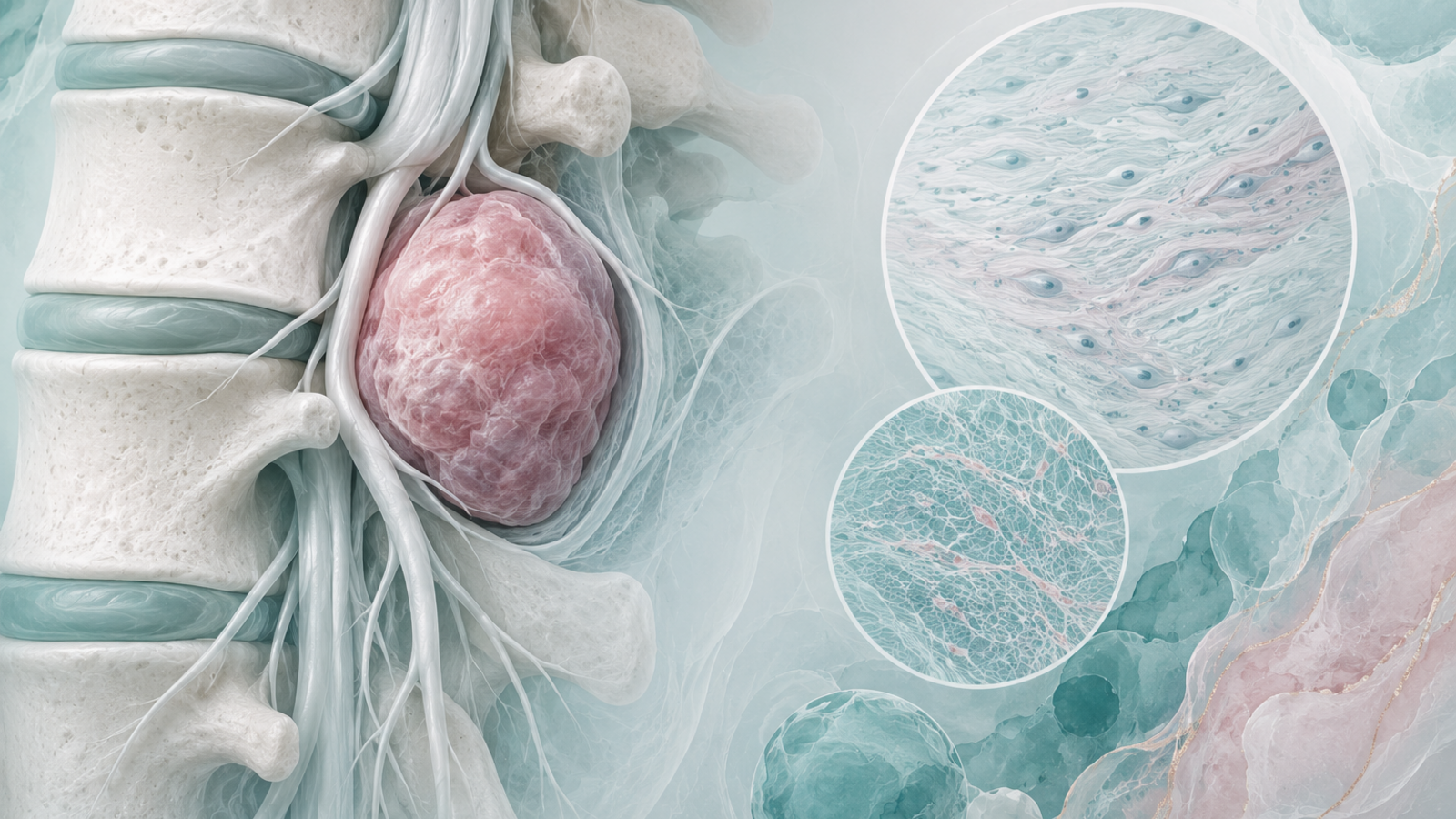
This article reviewed spinal myxomas and described two contrasting cases. It matters because spinal myxomas are rare and may resemble other spinal or paraspinal tumours on imaging.
Research snapshot
Article title: Spinal myxomas: review of a rare entity
Authors: Sabina Patel, Trisha Suji, Graeme Pang, Varinder S Alg, Ravindran Visagan, Zita Reisz, Jose P Lavrador, Ahilan Kailaya-Vasan, Gordan Grahovac
Publication type: Journal article
Publication date: 31 May 2022
Publication details: Journal of Surgical Case Reports. 2022;2022(5):rjac221.
PMID: 35665391
PMCID: PMC9156026
DOI: 10.1093/jscr/rjac221
Study type: Case series and literature review
Mr Grahovac’s involvement: Listed author on the publication
Original publication: View the original publication on PubMed
What this paper looked at
This paper looked at spinal myxomas, rare benign mesenchymal tumours that can occur in paraspinal tissues. The authors discussed two cases and reviewed diagnostic issues, including how imaging and histology help distinguish myxomas from other tumours.
Key points from the publication
The abstract reports two contrasting paraspinal cases and notes that histological analysis is the definitive method for diagnosis. Both patients had surgical resection, and follow-up imaging at six months showed no symptomatic or tumour recurrence in the reported cases.
Clinical relevance
The publication is relevant to differential diagnosis and surgical management of rare paraspinal tumours. It highlights the importance of histopathology when imaging appearances overlap with other tumour types.
What this means in context
Rare case-based evidence can inform clinical awareness, but it cannot predict outcomes for every patient. Management depends on symptoms, imaging, pathology, tumour behaviour, spinal stability and specialist multidisciplinary review.
View published abstract
The following abstract is reproduced from the original publication and is provided for reference. It may include technical terminology intended for clinical or academic audiences.
Intramuscular myxomas are rare, benign mesenchymal tumours, occurring predominantly in large skeletal muscles as large, slow-growing and painless masses. Spinal occurrence is rare, and may present incidentally, or diagnosed via localized symptoms secondary to local infiltration of surrounding structures. Differential diagnosis based on imaging includes sarcomas, meningiomas and lipomas. We discuss two contrasting cases presenting with well-circumscribed cystic paraspinal lesions indicative of an infiltrative tumour and discuss the radiological and histological differences that distinguish myxomas from similar tumours. Surgical resection of the tumour was performed in both cases, however one patient required surgical fixation due to bony erosion secondary to tumour infiltration. Immuno-histopathological analysis confirmed the diagnosis of a cellular myxoma. Follow up imaging at 6 months confirmed no symptomatic or tumour recurrence in both cases. Histological analysis is the definitive means for diagnosis to differentiate myxomas from other tumours. Recurrence is rare if full resection is achieved.
View the original publication on PubMed
About Mr Gordan Grahovac
Mr Gordan Grahovac is a Consultant Neurosurgeon and Complex Spinal Surgeon with expertise in managing complex spinal and neurosurgical conditions.
His work includes the assessment and treatment of patients with degenerative spinal conditions, spinal cord compression, spinal tumours, complex spinal pathology and conditions requiring specialist neurosurgical input.
His approach focuses on careful diagnosis, appropriate treatment planning and helping patients understand their options clearly.
Important note
This page is for educational purposes only and should not be taken as individual medical advice.
If you or someone you know has symptoms such as worsening headache, confusion, drowsiness, weakness, changes in speech, seizures, balance problems or symptoms following a head injury, seek urgent medical advice.
